CASE REPORT
Secondary Vulvar Hemorrhage After Ovariohysterectomy: Case Report of a Poorly Understood Complication
Colleen Guilfoyle1*, Set Sokol2 and Erin Katribe1
1Best Friends Animal Society, 5001 Angel Canyon Rd Kanab, UT, USA; 2Antech Diagnostics, Mars Petcare Science & Diagnostics, Loveland, CO, USA
Abstract
Secondary vulvar hemorrhage, also commonly referred to as uterine artery erosion, is a poorly understood postoperative complication of ovariohysterectomy. This report describes a case involving delayed vulvar hemorrhage beginning 6 days after an adult dog was spayed, culminating in fatal bleeding during a rescue procedure on day 12 post-operatively. A hemoabdomen was not present. Histopathology of the uterine stump revealed suppurative and chronic mural inflammation with vascular involvement. This is the first histopathologic description of this condition. The fatal outcome underscores the need for greater understanding of processes involved in delayed vulvar bleeding post-ovariohysterectomy. Additionally, early recognition and prompt surgical intervention may support better patient outcomes in suspected cases of secondary vulvar hemorrhage.
Keywords: ovariohysterectomy; secondary hemorrhage; histopathology; case report; postoperative complications
Citation: Journal of Shelter Medicine and Community Animal Health 2026, 5: 162 - http://dx.doi.org/10.56771/jsmcah.v5.162
Copyright: © 2026 C. Guilfoyle et al. This is an Open Access article distributed under the terms of the Creative Commons Attribution 4.0 International License (http://creativecommons.org/licenses/by/4.0/), allowing third parties to copy and redistribute the material in any medium or format and to remix, transform, and build upon the material for any purpose, even commercially, provided the original work is properly cited and states its license.
Received: 14 October 2025; Revised: 23 November 2025; Accepted: 23 November 2025; Published: 5 January 2026
Competing interests and funding: The authors declare no potential conflicts of interest. The authors have not received any funding or benefits from industry or elsewhere to conduct this study.
Correspondence: *Colleen Guilfoyle, Best Friends Animal Society, 5001 Angel Canyon Rd, Kanab, UT, USA. Email: colleeng@bestfriends.org
Reviewers: Sasha Nelson, Kirk Miller
Supplementary material: Supplementary material for this article can be accessed here.
Secondary vulvar hemorrhage, commonly known as uterine artery erosion, following ovariohysterectomy is a condition poorly described in veterinary literature. Serious, life-threatening bleeding 4–16 days after ovariohysterectomy has been reported by several authors and described in one case review article.1–3 Eleven cases of secondary vulvar hemorrhage were described in the 1973 review. This review introduced the terms ‘secondary hemorrhage’ and ‘uterine artery erosion’ for cases with a similar clinical presentation, but the pathogenesis of the clinical condition remains largely unknown. ‘Secondary hemorrhage’ is a term used in the human medical field to describe hemorrhage reported greater than 24 h postoperatively.4,5 Given the poor understanding of pathogenesis, secondary vulvar hemorrhage’ will be the term used in this report.
In some cases, vulvar bleeding begins intermittently and increases in severity over time while other cases present acutely. Severity can increase rapidly, becoming fatal within hours.1,3
Suggested ideas of how this occurs include erosion of the uterine vessels beneath the uterine body ligature.1,6 The cause of erosion is thought to be infection due to pyometra or breach in sterility, but this has not been confirmed by histopathology or culture.6 Another source proposes that a singular encircling ligature could cause more crushing and necrosis than a two-pass ligature, such as the miller’s knot.3
Despite anecdotal recognition among veterinarians performing high-volume sterilization surgeries, secondary hemorrhage remains poorly described in the literature. This report describes a case of fatal secondary vulvar hemorrhage. To our knowledge, this is the first report to include summarized surgical documentation, postmortem examination results, and histopathologic findings for this complication. Given the potential for rapid deterioration, awareness of this condition and further investigation into its pathogenesis are warranted.
Case
The patient was a 55-lb. (25 kg) female, intact, mixed-breed shelter dog estimated to be 2 years of age, who presented to a high quality, high volume spay-neuter (HQHVSN) clinic for an ovariohysterectomy during late-term pregnancy. No patient history was available aside from a recent transport from an animal control agency in eastern Arizona. Physical examination was unremarkable. An IDEXX SNAP 4DX was performed prior to anesthesia and was negative for Dirofilaria immitis antigen, as well as Anaplasma phagocytophilum, Anaplasma platys, Borrelia burgdorferi antibody, Ehrlichia canis, and Ehrlichia ewingii antibodies.
The patient was sedated with a 5 mg/mL injectable midazolam solution (generic) at 0.2 mg/kg intramuscularly (IM) and a 0.6 mg/mL injectable compounded buprenorphine solution (Wedgewood Pharmacy) at 0.02 mg/kg IM. An intravenous (IV) catheter was placed in the cephalic vein, and induction was achieved with a 10 mg/mL propofol solution (generic) at 2 mg/kg IV and a 100 mg/mL ketamine solution (generic) at 2 mg/kg IV. An additional 0.1 mg/mL dexmedetomidine solution (generic) was administered at 0.001 mg/kg IV intraoperatively. Intravenous Lactated Ringer’s solution was provided at 250 milliliters (mL) per hour during surgery. A 5 mg/mL meloxicam solution (Metacam, Boehringer Ingelheim) was administered subcutaneously at 0.04 mg/kg prior to surgery.
The patient was intubated and maintained on oxygen and isoflurane as needed. The surgical area was clipped and cleaned with a sterile 2% chlorhexidine scrub and solution.
A caudal umbilical midline incision was made and the right ovarian pedicle was isolated and ligated with Size-0 Monocryl then excised. This was repeated for the left ovarian pedicle. The uterine body was exteriorized, ligated with Size-0 Monocryl with a miller’s knot, circumferential knot, a miller’s knot, and two ligatures placed on each uterine vascular bundle. The uterine body was then excised. The linea was closed with 0 Monocryl in a simple continuous pattern. 0 Monocryl was used to close the subcutaneous layer in a simple continuous pattern. 0 Monocryl was used intradermally to close the skin. Cyanoacrylate was used, and an umbilical tattoo was applied. In addition, six skin staples were used along the length of the incision.
The patient awoke uneventfully from anesthesia and was discharged from the veterinary clinic at 46 h post-ovariohysterectomy. On day six post-ovariohysterectomy, the patient presented for vaginal hemorrhage (Fig. 1). No reports of vaginal discharge were reported prior to day 6. She was bright, alert, and responsive. Her physical examination was unremarkable aside from a consistent slow drip of bright red blood from her vulva. When the patient barked, larger amounts of frank blood and clotted blood were seen from her vulva. In-house laboratory findings revealed mild neutrophilia and mild hypochromic normocytic anemia (Table 1). AFAST (Abdominal focused assessment with sonography for trauma, triage, and tracking) was performed in-house and trace free fluid was identified. The uterine stump was identified on ultrasound with what appeared to be clotted tissue with no active bleeding, confirmed with color flow doppler.
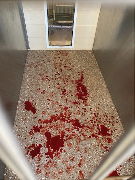
Fig. 1. Photograph of kennel the morning of day 6 post-ovariohysterectomy. No prior vaginal discharge was noted. Kennel compartments measured 1.6 m x 1.1 m.
The patient was hospitalized on days 6–8. Prescriptions were initiated on day 6 and included Yunnan Baiyao (Yunnan Baiyao Group Co., Ltd) at 1 capsule orally every 8 h, a maropitant citrate tablet (Cerenia, Zoetis) at 1.4 mg/kg orally once every 24 h, and an amoxicillin–clavulanate tablet (Clavamox, Zoetis) at 13.4 mg/kg orally every 12 h. An additional antibiotic was added on day 8 due to the white blood cell count continuing to rise, with an enrofloxacin tablet (Baytril, Elanco) administered at 13 mg/kg orally once every 24 h. The patient was discharged on day 8 due to no vulvar bleeding noted for 24 h. No additional vulvar bleeding was reported on days 8–11.
In-house complete blood count was performed on day 7, day 8, and day 12 (Table 1). Total white blood cell count increased at each recheck point. Total red blood cell count varied but remained below reference intervals for all recheck points.
The patient presented on day 12 post-ovariohysterectomy with vaginal hemorrhage, hypothermia (97°F [36.1°C]), and severe pallor (Figure 2). Blood pressure was unobtainable, suspected due to severe hypotension. A peripheral IV catheter was placed, and an LRS fluid bolus started. After initial 500 mL bolus, blood pressure was obtained with a mean arterial pressure of 45 millimeters (mm) of mercury (Hg). The patient was anesthetized and returned to surgery. Cardiopulmonary arrest occurred immediately upon return to surgery. Recovery attempts were unsuccessful.
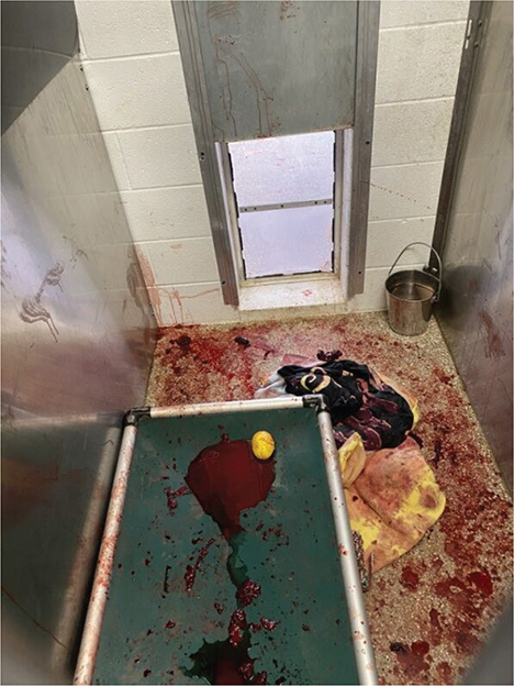
Fig. 2. Photograph of kennel the morning of day 12 post-ovariohysterectomy. Kennel compartments measured 1.6 m x 1.1 m.
A post-mortem examination was conducted immediately. No free fluid was present in the abdomen. The area between colon (dorsally) and bladder (ventrally) appeared granulomatous with many fibrous adhesions. Suture was palpable around a very short vaginal stump (cervical tissue palpable at most proximal aspect). The vaginal tissue had adhered to the ventral aspect of the colon, vaginal stump, and dorsal aspect of the bladder. One free encircling ligature was identified in the abdomen near the uterine stump.
Ovarian pedicles and uterine stump tissue were submitted for histopathologic review. The ovarian pedicles were consistent with granulomas expected with ovariohysterectomy surgery. The cervix was affected by suppurative mural inflammation. The mucosa was regionally intact with areas of erosion and lined by lymphoplasmacytic inflammation. Deeper in the muscular layer and adjacent adipose tissue were areas of chronic inflammation, with pockets of neutrophils and necrotic debris often lined by granulation tissue and large tortuous blood vessels.
Discussion
This case illustrates a rare but serious complication of ovariohysterectomy that has been sparsely documented in veterinary literature. Anecdotally, many veterinarians in high-volume settings are familiar with secondary vulvar hemorrhage as a possible complication.6 This is the first description of secondary vulvar hemorrhage following ovariohysterectomy that includes histopathologic description of the tissue.
Histopathology in this case suggests an inflammatory and possibly infectious process. Bacteria were not identified in the histopathologic specimens, but that does not rule out infection due to bacteria since histopathology is not a sensitive test for bacteria.7 Infection is a distinct possibility in this case given that this area was at a surgical site and near to a sutured mucosal orifice (cervix), both potential sources of bacterial infection. In addition, areas of tissue trauma, necrosis, and inflammation can be attractive substrates for any circulating infectious agents. Bacterial cultures were not collected in this case but should be considered in future investigations of secondary hemorrhage.
In this case, the complication was not recognized early, delaying intervention. In the existing literature describing this complication, successful early intervention via a second surgical procedure and ligation of the uterine stump or bleeding vessel is described.2,6
Given the multiple ligatures used in this initial surgery, this case questions the suggestion in the existing literature that secondary vulvar hemorrhage is the result of placing one encircling ligature around the uterine body.3 Additional questions remain in the HQHVSN community of how to prevent this postoperative condition. Ideas that have been proposed in the HQHVSN community include suture type and size, type and order of ligatures, and use of an ovariectomy (when appropriate) versus ovariohysterectomy.
It is unclear whether the dog’s gravid status contributed to this post-operative complication. Potential contributing factors include pregnancy-associated hemodynamic changes (such as increased uterine perfusion and vascular fragility) or rapid post-spay involution of the uterine stump that may have resulted in loosening of ligatures. Notably, previously published reports of secondary hemorrhage do not identify pregnancy as a documented risk factor.1,6 Further documentation and characterization of post-operative complications, including those occurring in pregnant patients, are needed to clarify whether gravid status influences the risk of secondary hemorrhage.
A limitation in this report is a lack of coagulation parameters, such as Prothrombin Time (PT) and Partial Thromboplastin Time (PTT). Diagnostics in this case were logistically limited to what could be performed in-house and given the number of clots found in the vulvar discharge, the patient’s ability to clot was assumed by the clinicians.
The name(s) of this collection of clinically similar cases introduces confusion. ‘Secondary vulvar hemorrhage’ is a term associated with childbirth in humans but is not commonly used in veterinary medicine.4,5 ‘Uterine artery erosion’ alludes to a hypothesis of pathogenesis, which may or may not be accurate.
This case highlights the importance of documenting post-operative complications in the literature as early intervention may have resulted in a different outcome. Further case reports and prospective studies are needed to clarify risk factors and mechanisms for secondary vulvar hemorrhage following ovariohysterectomy.
Conclusion
This case underscores that secondary hemorrhage after ovariohysterectomy, though uncommon, can progress rapidly and result in fatal outcomes. Early recognition of moderate to severe post-spay vaginal bleeding and timely return to surgery are critical to improving patient survival. This case is the first histopathologic description of secondary hemorrhage after ovariohysterectomy. Greater awareness among clinicians and further documentation of similar cases are needed to guide prevention and management of this complication.
Acknowledgments
The authors would like to acknowledge the clinicians who participated in this patient’s care and postmortem examination. Their work is supported by the dedication and skill of veterinary technicians, whose contributions are gratefully acknowledged. They would also like to acknowledge the frontline animal care staff who provide daily care for animals in shelters. Without their compassion and commitment, this work would not be possible. Additional thanks are given to Peter Wolf for ongoing collaboration and support in the editorial process.
References
| 1. | Pearson H. The Complications of Ovariohysterectomy in the Bitch. J Small Anim Pract. 1973;14(5):257–266. doi: 10.1111/j.1748-5827.1973.tb06457.x |
| 2. | Romagnoli S, Krekeler N, de Cramer K, Kutzler M, McCarthy R, Schaefer-Somi S. WSAVA Guidelines for the Control of Reproduction in Dogs and Cats. J Small Anim Pract. 2024;65(7):424–559. doi: 10.1111/jsap.13724 |
| 3. | Van Goethem B, Schaefers-Okkens A, Kirpensteijn J. Making a Rational Choice Between Ovariectomy and Ovariohysterectomy in the Dog: A Discussion of the Benefits of Either Technique. Vet Surg. 2006;35(2):136–143. doi: 10.1111/j.1532-950X.2006.00124.x |
| 4. | Paul PG, Prathap T, Kaur H, Shabnam K, Kandhari D, Chopade G. Secondary Hemorrhage After Total Laparoscopic Hysterectomy. JSLS. 2014;18(3):e2014.00139. doi: 10.4293/JSLS.2014.00139 |
| 5. | Pang T, Wu Z, Zeng H, Zhang X, Hu M, Cao L. Analysis of the Risk Factors for Secondary Hemorrhage after Abdominal Surgery. Front Surg. 2023;10:1091162. doi: 10.3389/fsurg.2023.1091162 |
| 6. | Bohling MW. Complications in Spay and Neuter Surgery. In: White S, ed. High-Quality, High-Volume Spay and Neuter and Other Shelter Surgeries. John Wiley & Sons, Ltd; 2020:339–361. doi: 10.1002/9781119646006.ch17 |
| 7. | Gupta E, Bhalla P, Khurana N, Singh T. Histopathology for the Diagnosis of Infectious Diseases. Indian J Med Microbiol. 2009;27(2):100–106. doi: 10.4103/0255-0857.49423 |