CASE REPORT
An Accessible Surgical Technique for Corneal Dermoid Removal as an Alternative to Enucleation: A Case Report
Sarah Flanders, DVM1, Matthew Chavkin, DVM, MS, DACVO2 and Brian DiGangi, DVM, MS, DABVP (Canine & Feline Practice, Shelter Medicine Practice)3
1Dumb Friends League, Denver, CO, United States; 2Mountain Star Veterinary Specialists, Greenwood Village, CO, United States; 3First Coast No More Homeless Pets, Jacksonville, FL, United States
Abstract
Corneal dermoids compromise ocular health and comfort. Dermoids are made up of cutaneous tissues, often including long hairs. When these hairs contact the cornea or other surrounding tissues, they can result in pain and discomfort. In resource-limited environments, veterinarians often perform enucleation of the affected eye to relieve the discomfort associated with corneal dermoids. This report describes a novel surgical technique to remove a canine corneal dermoid in a shelter using readily available equipment and basic surgical principles. Successful outcome supports an alternative to enucleation and/or referral for treatment of corneal dermoids. Although this procedure may result in decreased corneal clarity at the surgical site, the clinical outcome is acceptable and should be considered when access to specialized equipment and care is not feasible.
Keywords: ophthalmology; veterinary; corneal dermoidectomy; pseudopterygium; canine; animal welfare; case report
Citation: Journal of Shelter Medicine and Community Animal Health 2024, 3: 110 - http://dx.doi.org/10.56771/jsmcah.v3.110
Copyright: © 2024 Sarah Flanders et al. This is an Open Access article distributed under the terms of the Creative Commons Attribution 4.0 International License (http://creativecommons.org/licenses/by/4.0/), allowing third parties to copy and redistribute the material in any medium or format and to remix, transform, and build upon the material for any purpose, even commercially, provided the original work is properly cited and states its license.
Received: 28 July 2024; Revised: 12 September 2024; Accepted: 12 September 2024; Published: 21 November 2024
Reviewers: Staci Cannon, Sasha Nelson
Correspondence: Sarah Flanders, sarahlflanders@gmail.com
Supplementary material: Supplementary material for this article can be accessed here.
Competing interests and funding: No conflict of interests or funding to note.
Ocular dermoids are a type of choristoma, a mass of normal tissue present in an abnormal location. Dermoids often consist of keratinized epithelium, hair, blood vessels, fibrous tissue, fat, nerves, glands, smooth muscle, and cartilage. These benign growths occur during embryonic development and are characterized by histologically normal cutaneous tissue present in or around the eye. Frequently, the hairs of the dermoid contact the cornea, palpebral conjunctiva, and other surrounding tissues and cause corneal ulcers, keratitis, blepharospasm, and epiphora as a result.1 Superficial lamellar keratectomy, often with conjunctival graft, is the recommended treatment for corneal dermoids.2 This technique utilizes an operating microscope to aid in removing all of the dermoid tissue past the level of the outermost normal corneal epithelium, thus preventing tissue regrowth and recurrence of irritation and discomfort. This ophthalmic surgery requires specialized veterinary training, equipment, and follow-up care and is generally unavailable to animals within the sheltering system, in situations where finances are a concern, or when access to specialized skill and/or equipment is limited. To the author’s knowledge, there are no other published surgical treatment options for removal of corneal dermoids.
Animals in resource-limited environments, including within both shelter and community clinic settings, typically face decreased access to specialty care, mainly due to cost. The cost of superficial lamellar keratectomy is $3,000–$4,000 in the geographic region where this case was managed. In one report of US shelters, this is nearly twice the dollar amount available per animal in shelters with very high resource levels.3 Similarly, many private pet owners report barriers to obtaining non-emergency sick care with as much as 75% of these owners citing financial barriers.4 For veterinarians practicing incremental care, assessment of the skills/resources/equipment and the financial resources available to manage the case are two critical considerations in determining the ultimate care pathway.5 Although uncommon,1 corneal dermoids result in a decreased quality of life for the affected animal and must be treated appropriately. Many veterinarians opt for enucleation in order to relieve the discomfort associated with a corneal dermoid. In the 5 years prior to the case reported here, the author’s shelter, with an average annual intake of 20,000 animals, managed six dogs with corneal dermoids; three were adopted with medical waivers, two were enucleated, one was euthanized for unrelated conditions, and zero were referred for specialist care. Information available on shelter medicine specific social media boards6 confirms regular discussion of management techniques for shelter animals affected by corneal dermoids, including enucleation and few instances of experimental dermoid removal. This case report describes a surgical alternative to enucleation using a technique that is readily accessible to the general practitioner.
Case
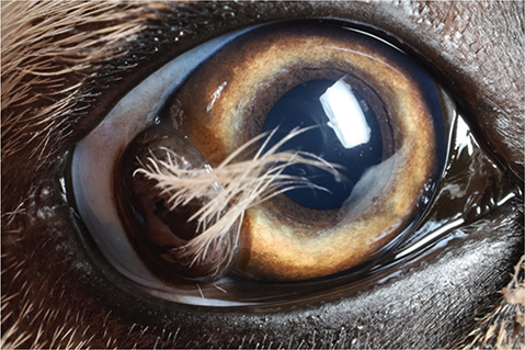
A 4-month-old intact female 16.0 kg mixed breed dog was relinquished to a private non-profit animal shelter in the western United States as part of an unintentional litter. No behavioral or medical history was provided. On physical examination, an approximately 6 mm × 8 mm corneal dermoid was present at the temporal limbus of the right eye from 8 to 10 o’clock. The corneal dermoid had many long hairs that were in constant contact with the central cornea (Fig. 1). As a result of the irritation, the dog was moderately blepharospastic, but had no other clinical signs. The rest of the physical examination was unremarkable, and no preoperative diagnostics were performed. Due to evidence of ongoing discomfort as a result of the dermoid, treatment options were considered and, ultimately, an experimental technique was attempted under the guidance of a local veterinary ophthalmologist who routinely provides consultation and ophthalmologic training to the shelter staff.

Fig. 1. Temporal limbal corneal dermoid of the right eye in a 4-month-old mixed breed dog.
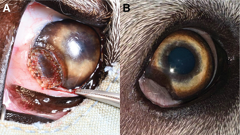
The dog was anesthetized with an intramuscular injectable combination of tiletamine and zolazepam (Tzed, Dechra) at 1.3 mg/kg, butorphanol (Torphadine, Dechra) at 0.07 mg/kg, and dexmedetomidine (Dexmedesed, Dechra) at 0.003 mg/kg and maintained on isoflurane throughout the procedure. The eye was aseptically prepared with a dilute betadine flush and draped routinely. The dermoid was sharply dissected away from the cornea using iris scissors, with care taken not to traumatize or perforate the associated corneal epithelium, the limbus, or the conjunctiva. Direct visualization was used to determine the depth of dissection and ensure removal of all pigmented skin-like dermoid tissue. Once removed, the tissue beneath consisted of mildly edematous central cornea with mild neovascularization and a circumferential raised margin of opaque tissue. The raised margin was cauterized with a Bovie low temperature handheld cautery to ensure complete destruction of the dermoid tissue (Fig. 2A). A temporary lateral partial tarsorrhaphy was performed with rapidly absorbed 5-0 Polyglactin 910 suture (Vicryl Rapide, Ethicon), ensuring coverage of the surgical site, to aid in healing, and to prevent trauma to the region. Total surgery time was 20 min.

Fig. 2. (A) Immediately postoperative corneal dermoidectomy (B) Four months postoperative pseudopterygium and corneal pigmentation.
Postoperative analgesia included carprofen (4.4 mg/kg PO q24h × 5d) and gabapentin (12.5 mg/kg PO BID × 5d). Gabapentin also aided in anxiolysis during the postoperative period. A topical antibacterial ointment (Neo-Poly-Bac, Bausch + Lomb) was applied twice daily for 7 days to prevent infection of the surgical site. An e-collar was placed for 10 days to prevent self-trauma to the surgical site. During the postoperative period, the dog was housed in a double compartment kennel along with one littermate of similar size. In accordance with shelter policy regarding age and vaccination status, the dog was not receiving regular walks or exercise time outside prior to surgery; this was not altered in the postoperative period. The total cost of the surgical procedure, supplies, and extended shelter length of stay (LOS) was $360.
A recheck was performed 2 days postoperatively, which showed granulation of the cornea where the dermoid was removed. Due to this region of the cornea being covered in adhered tissue, fluorescein staining was not performed. There was no evidence of discomfort (blepharospasm, pawing at the eye), infection, or additional inflammation (epiphora, discharge, conjunctival erythema, chemosis). At this time, the dog was cleared for adoption and adopted 4 days later. The prescribed medications were sent home with the adopter.
Two weeks and again 4 months postoperatively, the dog returned to the shelter for recheck appointments at no cost to the adopter. By 2 weeks postoperatively, a pseudopterygium had formed, wherein the conjunctiva spans over the cornea. At this time, the lateral tarsorrhaphy sutures had dissolved and were no longer present. By 4 months postoperatively, this pseudopterygium began to recede and became more translucent, indicating a successful healing process. The cornea where the pseudopterygium had previously been, and now receded, had mild dark brown pigmentation (Fig. 2B). Throughout the healing process, the dog did not exhibit any signs of discomfort or inflammation. No additional rechecks were warranted.
Discussion
In this case, a corneal dermoid was successfully removed in a shelter setting using readily available equipment and basic surgical principles, offering an alternative to enucleation when specialized ophthalmologic care was unavailable. This corneal dermoidectomy proved more cost and resource effective than enucleation, while also decreasing potential complications and allowing the patient to retain a comfortable, visual eye.
Surgical treatment of corneal dermoids using superficial or deep keratectomy and a conjunctival flap, is typically referred to veterinary ophthalmologists due to the specialized skill set and equipment needed. Superficial keratectomy requires an operating microscope to create an incision into the cornea in order to remove the entirety of the corneal dermoid.2 This surgical option is often not available to animals within a community clinic or shelter setting due to the costs of referral. If the dermoid is causing chronic irritation or impacting animal welfare, enucleation of the affected eye is often pursued.
Prior to surgery, three treatment options were available in this case. These included: benign neglect with waiver encouraging referral to a veterinary ophthalmologist, enucleation, or attempted removal. Direct specialist referral was not an option due to shelter budgetary constraints. Benign neglect with waiver was considered, though animals with medical waivers typically have a longer LOS than animals without waivers.7 Medical waivers imply a need for ongoing veterinary care and present a barrier to adoption; the average adopter may be unable and/or unwilling to put forth the finances and resources needed to care for a medical condition in an animal with whom they lack a bond. While waivers and counseling inform an adopter of the needs for ongoing medical care of an animal, they do not guarantee that the animal will receive the needed care after adoption. In cases where the animal’s welfare is compromised without the recommended medical intervention, alternatives need to be considered, as was done in this case.
A common surgical treatment option utilized to relieve the discomfort of corneal dermoids that can be performed by general practitioners and shelter veterinarians is enucleation. Enucleation was considered in this case, as it had been used to manage shelter dogs with corneal dermoids in this particular shelter in the past. A novel approach was ultimately decided on for this patient due to the availability of specialist guidance in hopes of sparing an otherwise healthy, comfortable, and visual eye.
The surgical procedure reported here, corneal dermoidectomy, has advantages over benign neglect, enucleation, and ophthalmology referral for superficial lamellar keratectomy (Table 1). Firstly, this technique costs less than both referral and enucleation (Supplementary Material). The total cost of care for the corneal dermoidectomy, including the surgery, extended shelter LOS, medications, and supplies was approximately $360. A key component of corneal dermoidectomy is the use of cautery to epilate any remaining follicular tissue, ensuring complete destruction of the hairs. The cautery used here is inexpensive (approximately $50) and readily accessible to the general practitioner. In contrast, the total cost of an enucleation surgery within the shelter setting, including extended shelter LOS, medications, and supplies is approximately $460. The cost of superficial keratectomy and enucleation by local veterinary ophthalmologists is approximately $4,000 and $3,000, respectively. Secondly corneal dermoidectomy required less time under anesthesia than enucleation and totaled about the same time as superficial keratectomy. Within the shelter setting, both corneal dermoidectomy and enucleation are comparable in that both require extended LOS and postoperative medications. However, this technique required less surgical equipment than enucleation and the animal was cleared for adoption faster than if referral were pursued.
The aim of this technique is to remove dermoid tissue without removing corneal tissue. In contrast, the aim of superficial lamellar keratectomy is the intentional removal of corneal epithelium, using an operating microscope to make a 30% depth incision into the cornea itself. For this reason, a key limitation of this technique is the reliance of visual cues and clinician discretion in determining if complete dermoid removal has been achieved. Although not performed in this case, the use of fluorescein staining may be used immediately postoperatively to determine if corneal epithelium remains intact. In addition, fluorescein staining was not performed at the initial physical exam due to the absence of clinical signs suggestive of a corneal ulcer (corneal edema, neovascularization, discharge) and clinician discretion, but may be helpful in cases where corneal ulceration is suspected. Staining was not used during postoperative rechecks as the corneal epithelium remained covered with conjunctiva.
Additionally, corneal dermoidectomy offers a more positive welfare outcome for the animal than either benign neglect or enucleation. In reference to the Five Domains, this approach most directly positively impacts the domains of Health and Mental State offering high functionality of the affected eye and the absence of pain.8 Especially in cases with long hairs contacting the cornea, corneal dermoids provide a constant source of irritation to the cornea and surrounding structures, frequently resulting in corneal ulceration.1 The discomfort of corneal dermoids is evident clinically; affected animals are blepharospastic, frequently paw at their faces and eyes, and have ocular discharge. Most dogs are diagnosed with corneal dermoids between 1 and 2 years of age,1 resulting in prolonged and chronic discomfort. For these reasons, benign neglect leaves an animal in a chronic negative welfare state. Although removing the source of discomfort, enucleation results in decreased overall vision and results in a functional impairment. Many animals may be startled when approached on the side of the enucleated eye or have trouble navigating their environment after enucleation surgery.9 Any future trauma or disease of the remaining eye can reduce or eliminate vision altogether.
Corneal dermoidectomy has fewer potential complications than enucleation surgery, which typically include surgical site infection, hemorrhage, contralateral blindness, and retention of adnexal remnants,10 but poses similar risks to superficial lamellar keratectomy, which include infection, recurrence of the lesion, corneal pigmentation, and corneal perforation. Both dermoid removal surgeries provide a faster return to activity (7 days) when compared to enucleation surgery (14 days). There were no surgical complications associated with this case of dermoid excision. The outcome of using this technique to remove a central corneal dermoid, not associated with the limbus, is expected to be similar. The pseudopterygium seen during healing in this case was an expected result; conjunctivalization indicates that there was an absence of limbal stem cells in the region of the dermoid, allowing conjunctival epithelial cells to migrate onto the corneal surface postoperatively. This common finding essentially creates a conjunctival graft to aid in the healing process. Conjunctivalization of the cornea can occur as the result of any corneal trauma, infection, or immunologic disease11 and is not unique in this case. Due to the location of the pseudopterygium outside of the visual axis in this case, corneal clarity is not a concern and overall vision will not be affected.12 However, based on the known corneal healing process, the pseudopterygium is expected to continue to recede.2 Long-term, adequate comfort and function of the eye will be achieved.
In addition to canines, corneal dermoids have been documented in felines, birds, lagomorphs, rodents, equids, bovine, and camelids. Results of this technique if utilized in other species are unknown, but expected to be similar.
Conclusion
The removal of irritating corneal dermoids promotes positive welfare of the affected animal by removing a source of discomfort to the cornea and surrounding ocular structures and preserving normal ocular function. When specialist referral is not an option, many general and shelter practitioners resort to enucleation. By electing to perform corneal dermoidectomy in lieu of enucleation in a shelter setting, a functional and visible eye was spared. The surgery did not require more shelter costs or resources when compared with enucleation surgery and did not have any associated complications. This surgical approach has not previously been reported in the scientific literature and describes an acceptable alternative to enucleation that can be performed by general and shelter practitioners with readily accessible resources.
Author contributions
Sarah Flanders: Conceptualization, Investigation, Methodology, Writing – Original Draft.
Brian DiGangi: Writing – Review and Editing.
Matthew Chavkin: Writing – Review and Editing.
Acknowledgments
Acknowledgments to Dumb Friends League, a private non-profit animal shelter who provided animal care, surgical and medical material, and financial resources as well as to Mountain Star Veterinary Specialists who provided surgical mentorship and supplies.
Author notes
The material in this manuscript has not been included elsewhere.
References
| 1. | Badanes Z, Ledbetter EC. Ocular dermoids in dogs: a retrospective study. Vet Ophthalmol. 2019;22:760–766. doi: 10.1111/vop.12647 |
| 2. | Gelatt KN, Ben-Shlomo G, Gilger BC, Kern TJ, Plummer CE. Veterinary Ophthalmology. Hoboken, NJ: John Wiley & Sons; 2021. |
| 3. | Gunter LM, Gilchrist RJ, Blade EM, et al. Emergency fostering of dogs from animal shelters during the COVID-19 pandemic: Shelter practices, foster caregiver engagement, and dog outcomes. Front Vet Sci. 2022;9:862590. doi: 10.3389/fvets.2022.862590 |
| 4. | Wiltzius AJ, Blackwell MJ, Krebsbach SB, et al. Access to Veterinary Care: Barriers, Current Practices, and Public Policy. 2018. https://pphe.utk.edu/wp-content/uploads/2020/09/avcc-report.pdf. Accessed June 4, 2024. |
| 5. | Program for Pet Health Equity Center for Behavioral Health Research. Incremental Veterinary Care Guide. 2023. https://pphe.utk.edu/wp-content/uploads/2023/03/AlignCare-Incremental-Care-Guide.pdf. Accessed June 4, 2024. |
| 6. | Shelter Medicine Veterinarians Facebook Group. Examples of Shelter Veterinarians and their Searches for Alternatives to Enucleation and Experimental Techniques. https://www.facebook.com/groups/186499308373730/search/?q=dermoid. Accessed July 19 2024. |
| 7. | Pockett J, Orr B, Hall E, Chong WL, Westman M. Investigating the impact of indemnity waivers on the length of stay of cats at an Australian Shelter. Animals. 2019;9(2):50. doi: 10.3390/ani9020050 |
| 8. | Mellor DJ, Beausoleil NJ, Littlewood KE, et al. The 2020 five domains model: including human–animal interactions in assessments of animal welfare. Animals. 2020;10(10):1870. doi: 10.3390/ani10101870 |
| 9. | Veterinary Information Network. Describes Physical Limitations Associated with Enucleations. https://veterinarypartner.vin.com/default.aspx?pid=19239&id=4951449. Accessed July 25, 2024. |
| 10. | Ward AA, Neaderland MH. Complications from residual adnexal structures following enucleation in three dogs. J Am Vet Med Assoc. 2011;239(12):1580–1583. doi: 10.2460/javma.239.12.1580 |
| 11. | Joussen AM, Poulaki V, Mitsiades N, et al. VEGF-dependent conjunctivalization of the corneal surface. Investig Ophthalmol Vis Sci. 2003;44(1):117. doi: 10.1167/iovs.01-1277 |
| 12. | Gelatt KN. Wsava state of the art lecture. J Small Anim Pract. 1997;38(8):328–335. doi: 10.1111/j.1748-5827.1997.tb03479.x |